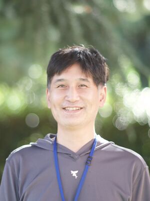
Takeo Saneyoshi
Bibliography
- 1996 Graduated with a degree in Animal Science, Faculty of Agriculture, Hokkaido University.
- 1998 M.S. Graduate School of Agricultural and Life Sciences, The University of Tokyo
- 2002 Ph.D. Graduate School of Medicine, The University of Tokyo. 2002 (Prof. Katsuhiko Mikoshiba)
- 2002-2007 Post-doc. Vollum Institute, Oregon Health & Science University (Prof. Thomas Soderling)
- 2007-2009 Post-doc. National Institute of Advanced Industrial Science and Technology (Dr. Toru Natsume)
- 2009-2016 Post-doc. Brain Science Institute, RIKEN (Dr. Yasunori Hayashi)
- 2016-present. Associate Professor. Dept. Pharmacology, Kyoto University, Graduate School of Medicine (Prof. Yasunori Hayashi)
Research topics
Dorsoventral axis formation regulated by calcium signaling in the early embryo
We screened several calcium-dependent enzymes to identify downstream molecules of the inositol 3-phosphate-Ca2+ pathway, which acts as a ventralizing signal during dorsoventral axis formation during early development, and found that the ventralizing signal is mediated by NFAT, a transcription factor regulated by the protein phosphatase calcineurin. When NFAT was repressed in the ventral region, the Wnt-β-catenin pathway of the classical Wnt pathway was activated and the fate was converted from ventral to dorsal. In contrast, the Wnt-Ca2+ pathway, which acts as a ventralizing signal, activated the calcineurin-NFAT pathway. These results suggest that the default fate of the early developing embryo is dorsal, and the transcripts of the calcineurin-NFAT pathway by local calcium signaling generates dorsoventral polarity and form the dorsoventral axis[1][2][3].
Neuronal activity-dependent neuronal morphogenesis
Neurons are highly polarized cells. Along with developmental stages, neurons acquire characteristic structures: axons, dendrites, and dendritic spines. In hippocampal neurons, neural activity promotes dendritic arborization and subsequently to form dendritic spines. In this process, the CaMKI-CaMKK cascade causes activation of the transcription factor CREB via the ERK pathway, and its transcript Wnt2 causes dendrite morphogenesis[4].
Using proteomic analysis of calmodulin kinase binding proteins, we found that CaMKK binds to βPIX, a guanine nucleotide exchange factor (GEF). In neurons, this signaling complex allows CaMKK and its downstream kinase CaMKI to phosphorylate Ser-516 of βPIX and promote GEF activity. Furthermore, we found that activity-dependent spine formation is regulated via the CaMKK-βPIX signaling complex, which is activated by glutamate receptors (NMDARs) [5].
We also found that a membrane-localized CaMKI&gamma: isoform acts downstream of TRPC5 channels to regulate axon formation and elongation[6](J. Neurosci., 2009), and the CaMKK/CaMKI/βPIX/GIT1 signaling complex regulates neural migration of cerebellar granule cells and medulloblastoma cell lines via Rac1 signaling[7].
In this series of findings, we demonstrated the biological significance of CaMKI, which was previously known as an orphan enzyme whose function was unknown, and in particular that it is an extremely important enzyme that converts calcium signals into biochemical reactions during neurogenesis[8]. CaMKI is also a molecule that links calcium to the ERK pathway in synaptic plasticity[9], and is expected to function not only during development but also in mature neurons.
Molecular mechanisms of synaptic plasticity (2009-present)
Synapses, the sites of neuronal communication, change their efficiency of transmission depending on the stimulus received. Synaptic plasticity is thought to be the basis of memory. There is a positive correlation between the size synapse and strength of synaptic transmission, and the actin cytoskeleton is responsible for structural changes[10][11].
We focused on the morphological changes of synapses associated with long-term potentiation (structural LTP) and analyzed the dynamics of synaptic proteins by live-imaging. We found that cofilin1, a regulator of the actin skeleton, rapidly enters the spine after stimulation and increases its synaptic concentration, stabilizing actin fibers and spine structure. We also found that postsynaptic densities (PSDs) do not change immediately after stimulation, but increases in a protein synthesis-dependent manner about one hour later[12].
A major question in memory research is how to store a transient synaptic stimuli as long-term information, and we found that CaMKII and the Rac-guanine nucleotide exchange factor Tiam1 form a persistent and stable signaling complex at LTP-stimulated synapses. Because the two molecules activated each other and remained active for a long-time, we named this mode of activation the reciprocally activating kinase-effector complex (RAKEC), which can convert a transient increase in calcium concentration into long-lasting Rac1 activity and synaptic structures [13][14].
To extend our findings from the cellular level to the individual level, we generated knock-in mice with CaMKII-mediated RAKEC formation. The knock-in mice with RAKEC deficient mutations in Tiam1 or CaMKIIα showed almost complete loss of synaptic plasticity in hippocampal slices. These results suggest that RAKEC formation is a key component of learning. Furthermore, the formation of long-term memory in the novel object recognition test was impaired (ref [15] and unpublished data). Thus, RAKECs made by Tiam1 and CaMKIIα may be involved in the systems-level processes that lead to the formation of long-term memory.
In addition to the above studies at the cellular level, we are also examining the mechanism of long-term memory at the molecular level. Since proteins are replaced by turnover in cells, lifelong memory requires a mechanism for long-term maintenance of “activity of memory molecule” in the synapse. Liquid-liquid phase-separation, which has attracted attention as a membrane-free organelle, is one of the mechanisms that maintain the compartmentalization of substances in cells. One of the conditions for phase separation is the ability to form multimers of protein. It is thought that CaMKII, a dodecamer, serves as the core of phase separation, accumulating and concentrating functional molecules and contributing to long-term memory[14]. In fact, in experiments using purified proteins, CaMKII is phase-separated from synaptic proteins (unpublished). We are testing the hypothesis that CaMKII functions as a “memory molecule” together with the aforementioned RAKEC.
In the future, we would like to extend and develop this research to answer the question, “Why are biological materials replaced but memories are not?”
Publications
- ↑
Saneyoshi, T., Kume, S., Natsume, T., & Mikoshiba, K. (2000).
Molecular cloning and expression profile of Xenopus calcineurin A subunit(1). Biochimica et biophysica acta, 1499(1-2), 164-170. [PubMed:11118649] [WorldCat] [DOI] - ↑
Saneyoshi, T., Kume, S., Amasaki, Y., & Mikoshiba, K. (2002).
The Wnt/calcium pathway activates NF-AT and promotes ventral cell fate in Xenopus embryos. Nature, 417(6886), 295-9. [PubMed:12015605] [WorldCat] [DOI] - ↑
Saneyoshi, T., Kume, S., & Mikoshiba, K. (2003).
Calcium/calmodulin-dependent protein kinase I in Xenopus laevis. Comparative biochemistry and physiology. Part B, Biochemistry & molecular biology, 134(3), 499-507. [PubMed:12628380] [WorldCat] [DOI] - ↑
Wayman, G.A., Impey, S., Marks, D., Saneyoshi, T., Grant, W.F., Derkach, V., & Soderling, T.R. (2006).
Activity-dependent dendritic arborization mediated by CaM-kinase I activation and enhanced CREB-dependent transcription of Wnt-2. Neuron, 50(6), 897-909. [PubMed:16772171] [WorldCat] [DOI] - ↑
Saneyoshi, T., Wayman, G., Fortin, D., Davare, M., Hoshi, N., Nozaki, N., Natsume, T., & Soderling, T.R. (2008).
Activity-dependent synaptogenesis: regulation by a CaM-kinase kinase/CaM-kinase I/betaPIX signaling complex. Neuron, 57(1), 94-107. [PubMed:18184567] [PMC] [WorldCat] [DOI] - ↑
Davare, M.A., Fortin, D.A., Saneyoshi, T., Nygaard, S., Kaech, S., Banker, G., Soderling, T.R., & Wayman, G.A. (2009).
Transient receptor potential canonical 5 channels activate Ca2+/calmodulin kinase Igamma to promote axon formation in hippocampal neurons. The Journal of neuroscience : the official journal of the Society for Neuroscience, 29(31), 9794-808. [PubMed:19657032] [PMC] [WorldCat] [DOI] - ↑
Davare, M.A., Saneyoshi, T., & Soderling, T.R. (2011).
Calmodulin-kinases regulate basal and estrogen stimulated medulloblastoma migration via Rac1. Journal of neuro-oncology, 104(1), 65-82. [PubMed:21107644] [WorldCat] [DOI] - ↑
Saneyoshi, T., Fortin, D.A., & Soderling, T.R. (2010).
Regulation of spine and synapse formation by activity-dependent intracellular signaling pathways. Current opinion in neurobiology, 20(1), 108-15. [PubMed:19896363] [PMC] [WorldCat] [DOI] - ↑
Schmitt, J.M., Guire, E.S., Saneyoshi, T., & Soderling, T.R. (2005).
Calmodulin-dependent kinase kinase/calmodulin kinase I activity gates extracellular-regulated kinase-dependent long-term potentiation. The Journal of neuroscience : the official journal of the Society for Neuroscience, 25(5), 1281-90. [PubMed:15689566] [PMC] [WorldCat] [DOI] - ↑
Saneyoshi, T., & Hayashi, Y. (2012).
The Ca2+ and Rho GTPase signaling pathways underlying activity-dependent actin remodeling at dendritic spines. Cytoskeleton (Hoboken, N.J.), 69(8), 545-54. [PubMed:22566410] [WorldCat] [DOI] - ↑
Kim, K., Saneyoshi, T., Hosokawa, T., Okamoto, K., & Hayashi, Y. (2016).
Interplay of enzymatic and structural functions of CaMKII in long-term potentiation. Journal of neurochemistry, 139(6), 959-972. [PubMed:27207106] [WorldCat] [DOI] - ↑
Bosch, M., Castro, J., Saneyoshi, T., Matsuno, H., Sur, M., & Hayashi, Y. (2014).
Structural and molecular remodeling of dendritic spine substructures during long-term potentiation. Neuron, 82(2), 444-59. [PubMed:24742465] [PMC] [WorldCat] [DOI] - ↑
Saneyoshi, T., Matsuno, H., Suzuki, A., Murakoshi, H., Hedrick, N.G., Agnello, E., O'Connell, R., Stratton, M.M., Yasuda, R., & Hayashi, Y. (2019).
Reciprocal Activation within a Kinase-Effector Complex Underlying Persistence of Structural LTP. Neuron, 102(6), 1199-1210.e6. [PubMed:31078368] [PMC] [WorldCat] [DOI] - ↑ 14.0 14.1
Saneyoshi, T. (2021).
Reciprocal activation within a kinase effector complex: A mechanism for the persistence of molecular memory. Brain research bulletin, 170, 58-64. [PubMed:33556559] [WorldCat] [DOI] - ↑
Kojima, H., Rosendale, M., Sugiyama, Y., Hayashi, M., Horiguchi, Y., Yoshihara, T., Ikegaya, Y., Saneyoshi, T., & Hayashi, Y. (2019).
The role of CaMKII-Tiam1 complex on learning and memory. Neurobiology of learning and memory, 166, 107070. [PubMed:31445077] [WorldCat] [DOI]
Teaching experience
- B11a/b Pharmacology lecture and practice
Academic Society
- Society for Neuroscience
- The Japanese Society for Neuroscience
- The Japanese Society for Neurochemistry
Personal Aspects
Hobbies:
Fishing (especially small freshwater fishing and salt lures), playing the acoustic guitar, baking cakes
Reading (modern and contemporary Japanese history)
Contact address
Konoe-cho, Yoshida, Sakyo-ku, Kyoto 606-8501, Japan
Room 404, Building A, Department of pharmacology, Graduate School of Medicine, Kyoto University
E-mail address: saneyoshi.takeo.3v@kyoto-u.ac.jp
Tel: 075-753-4393

 https://orcid.org/0000-0003-2361-3339